Baby’s first breaths of life captured for the first time
By Hudson Institute communications
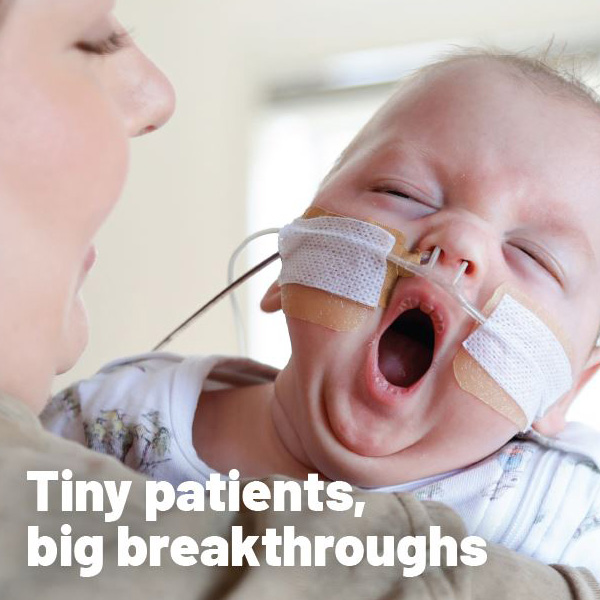
For the first time, doctors and researchers have captured moving ultrasound images of the lungs of newborn babies as they take their first breaths.

The world-first research, involving Hudson Institute of Medical Research, Monash University and the Royal Women’s Hospital, is a breakthrough in understanding how human lungs transition from the womb to taking the first breaths at birth.
Researchers say the new information could lead to the diagnosis of severe breathing problems in very preterm babies in first minutes of life, instead of the current several hours, allowing for life-saving treatment.
In the DOLFIN study researchers captured images of 115 newborn lungs immediately after birth at the Women’s. This included images of 28 full-term babies before they took their first breaths after birth. The research has been published in Resuscitation and the BMJ’s Archives of Disease in Childhood.
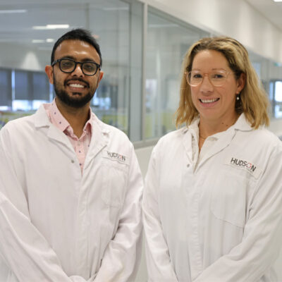
“When looking at the lungs immediately after birth, they look more like liver and are filled with liquid, but as they fill with air for the first time, the liquid is pushed out and the lungs open as they aerate,” lead researcher Dr Douglas Blank from the Women’s, Hudson Institute and Monash University said.
“Within 10 minutes, the lungs of these healthy babies appear to have nearly completed the adjustment needed after birth, although it does take, on average, up to four hours for all the liquid to be removed from the baby’s lungs.
“This research has important implications because the quality of the imaging and speed at which babies transition to normal lungs suggests we can make a diagnosis of lung problems in newborn babies within the first 20 minutes after birth using ultrasound. This is particularly important for premature infants as 50 per cent have severe breathing problems and require intensive treatment. Currently it takes several hours to assess the severity of breathing problems.”
Dr Blank is leading a new study, called DOLFIN Jr, to test whether it is possible to use imaging of the lungs in the first minutes of life to accurately predict which extremely pre-term babies (born before 29 weeks) need intensive breathing support and which babies will benefit from minimal breathing support.
Premature babies with severe breathing problems require lifesaving medication called surfactant. It is administered by placing a breathing tube into this tiny baby’s airway and putting the baby on a ventilator which is a machine that helps them breathes. This process can cause long-term damage to the fragile lungs so should only be done if the baby has severe breathing problems.
Babies without severe breathing problems do not need ventilation and can receive CPAP breathing assistance which decreases the risk of lung damage and death.
The window to decide which respiratory treatment is best suited for the baby, is small.
Currently, extremely preterm babies receive minimal breathing support with CPAP to start. If their oxygen levels are too low, indicating they are struggling to breathe they are given surfactant and put onto a breathing machine.
“Ultimately, we aim for babies who need the surfactant and help via a breathing machine to receive that support much earlier, while those whose lungs are functioning better, will receive minimal breathing support,” Dr Blank said. “Lung ultrasound may be able to tell us which approach is suited for each baby. Targeting breathing support more accurately will reduce the risk of lung disease and death in both groups,”
The Women’s Director of Newborn Research Professor Peter Davis said the new research represented a breakthrough in understanding human lungs in transition at birth.
“We really didn’t understand what was happening in those first seconds of birth as the only way to do it would be to x-ray a baby every few seconds – something that is not beneficial to either mum or baby. But the ultrasound has allowed us to gain an insight and see what the lungs are doing.
“With this technology, we can now look at very preterm babies immediately after birth and see which ones are going to develop lung disease and given them the extra help of mechanical ventilation and surfactant,” Professor Davis said.
The new DOLFIN Jr study is being funded initially by a US$50,000 General Electric grant and the Emergency Medicine Foundation and is being undertaken at the Royal Women’s Hospital and the Monash Medical Centre.
Background
115 full term babies were involved in the study with ultrasound used to image their breathing in the first minutes after birth. The very first breath of 28 babies was captured on ultrasound with another 35 within the first four breaths.
Surfactant is a naturally produced substance, a kind of foamy, fatty liquid that acts like grease within the lungs. Without it, the air sacs open but have difficulty remaining open because they stick together. Surfactant usually appears in the fetus’ lungs at about the 24th week of pregnancy and gradually builds up to its full level by the 37th week. If a premature baby is lacking surfactant, artificial surfactant may be given.
Contact us
Hudson Institute communications
t: + 61 3 8572 2697
e: communications@hudson.org.au
About Hudson Institute
Hudson Institute’ s research programs deliver in three areas of medical need – inflammation, cancer, women’s and newborn health. More
Hudson News
Get the inside view on discoveries and patient stories
“Thank you Hudson Institute researchers. Your work brings such hope to all women with ovarian cancer knowing that potentially women in the future won't have to go through what we have!”


